
 Neurobiology
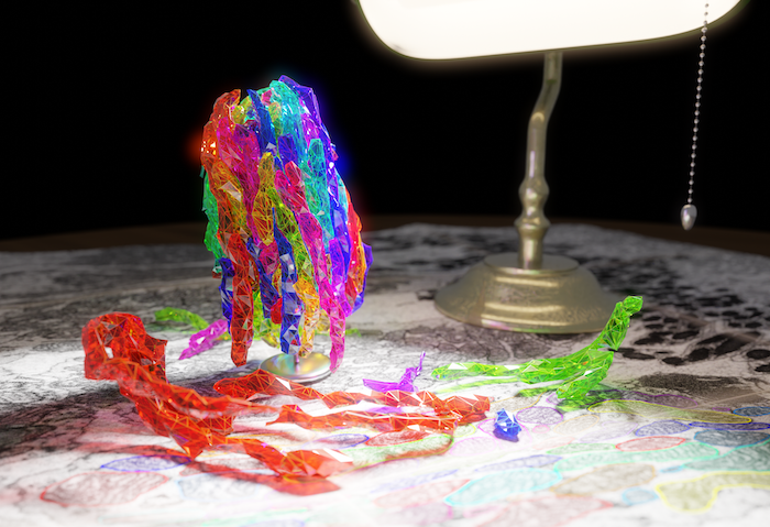
Mitochondria as microlenses in the eye – the evolution of an improved camera sensor
The neurons of the eye that detect light must be very sensitive. They also need a lot of energy, so they contain many mitochondria - however, mitochondria scatter light. In our recent study, we show how evolution may have found a way to have the best of both worlds: Why not build mitochondria into a lens that helps, rather than hurts, visual sensitivity?
The back of the eye is lined with a layer of specialized light-sensitive neurons in the retina, arranged in a mosaic, called photoreceptors. Each photoreceptor has a tapered elongated shape—like a bottle—oriented perpendicular to the retina and pointing toward the pupil of the eye. The narrow end, which points away from the pupil and contains the molecules that convert light into an electrical signal, is called the outer segment.
From past research, we know that each photoreceptor acts as a miniature antenna: it receives photons and guides them toward the outer segment in the same way that lenses bend light. This phenomenon improves the sharpness of our vision: ordinarily, due to light diffraction by the lens of the eye, such small photodetectors would see a small amount of the light intended for their neighbors, blurring the image we see. The shape of the photoreceptor rejects this stray light, sharpening our vision. This property is important for diagnostic tools used by ophthalmologists (medical doctors who specialize in diseases of the eye) to view light reflected from the retina, even allowing the observation of individual photoreceptors.
However, this picture of the optical nature of photoreceptors is incomplete. Photoreceptors are not uniformly transparent like glass—like other cells, they contain many organelles that are refractive, so they scatter light. In particular, mitochondria are highly refractive. Mitochondria are the cellular power plant, and photoreceptors need many to supply energy for the constant conversion of light energy into electrical signals. In theory, mitochondria could be dispersed evenly throughout the cell; however, photoreceptor mitochondria are long and packed into a tight bundle next to the outer segment; as a result, these mitochondria are the last obstacle through which light must pass before reaching its target.
This structure intrigued us. Could this arrangement of mitochondria reduce light scatter?
We study a fascinating animal—the thirteen-lined ground squirrel—that hibernates in the winter, and during this time, its photoreceptor mitochondria change: they become less elongated and more tangled. If our hypothesis was true, light passage through photoreceptors in the hibernating animal should be worse.
We tested this idea in two ways. First, we built a computer program to simulate the flow of light through photoreceptors. Second, we prepared retinal samples in which photoreceptors were separated from the remainder of the retina to test them in real life. We illuminated these samples from below, mimicking the path taken by light in the eye, and captured 3D images of the light passing through intact photoreceptors. This experiment would not have been possible without the special cellular properties that ground squirrels have that allow them to safely hibernate, keeping them healthy in harsh conditions that humans cannot survive.
Surprisingly, we found that instead of scattering light, mitochondria greatly increased the intensity of light passing through them. And yes, this effect was indeed about 20% worse in samples from hibernating squirrels. Also, the focused beam of light perfectly overlapped the shape of the outer segment, which should optimize capture of photons. We also showed in simulations that photoreceptors without mitochondria could also focus light, but less tightly, resulting in a beam of light that does not overlap the outer segment.
We found one more fascinating by-product of mitochondria-based focusing. Under the microscope, photoreceptors that were slightly tilted nevertheless focused light. However, we realized that the tilt of the photoreceptor would prevent the focused beam from overlapping with the outer segment. This effect matches a phenomenon first observed nearly 100 years ago in human vision, known as the Stiles-Crawford effect. We believe this means we are observing a mechanism that has evolved to increase sensitivity and shield diffracted light, ingeniously using the mitochondria that are already needed for energy production. Interestingly, many non-mammals (especially birds) have evolved a separate organelle in this exact location—an oil droplet—to serve such an optical role.
This long-established evolutionary invention has only recently been rediscovered by humans. In digital cameras, a single large lens focuses light onto a sensor array where tiny detectors encode the pattern of light with electrical signals. As with our eye, miniaturization of these photodetectors results in blur from diffraction. However, recent camera advances have offered a now-familiar solution: tiny lenses in front of each detector that shield it from unwanted light, increasing sharpness.
Importantly, many widely used diagnostics in research and in medicine depend upon measurements of the reflection of light from the retina, and the most innovative techniques exploit light guidance by photoreceptors. However, the effect of mitochondria on these results is still unclear. Because mitochondria dysfunction contributes to retinal diseases, if we can harness an improved knowledge of their optical characteristics to better understand such diagnostics, we may be able to detect retinal diseases and intervene earlier.
Original Article:
Ball, J. M., Chen, S., Li, W. (2022). Mitochondria in cone photoreceptors act as microlenses to enhance photon delivery and confer directional sensitivity to light. Science Advances, 8(9). https://doi.org/10.1126/sciadv.abn2070Next read: More than meets the eye: the histones revealed as enzymes by Oscar A. Campos , Siavash K. Kurdistani
Edited by:
Dr. Ilaria Di Meglio , Senior Scientific Editor
We thought you might like
Living without mitochondria: the downfall of one textbook truth
Oct 3, 2016 in Evolution & Behaviour | 3.5 min read by Lukáš NovákExercise helps restore aged muscles
Apr 20, 2018 in Health & Physiology | 3.5 min read by Matthew Robinson , Sreekumaran NairThe mystery of mistletoe mitochondria
Dec 21, 2018 in Evolution & Behaviour | 4 min read by Andrew Maclean , Janneke Balk , Etienne MeyerPowering up the emergency response to infections
Jun 4, 2020 in Health & Physiology | 3 min read by Jayna J. Mistry , Kristian M. Bowles , Stuart A. RushworthMore from Neurobiology
Can we use a magnet to see brain inflammation?
Sep 25, 2023 in Neurobiology | 4 min read by Raquel Garcia-Hernandez , Santiago Canals , Silvia de SantisSurprising Behavior Changes in Genetically Modified Syrian Hamsters
Aug 30, 2023 in Neurobiology | 4 min read by Susan Lee , Kim Huhman , Jack TaylorTo achieve goals, we definitively need our neurons
Mar 10, 2023 in Neurobiology | 3.5 min read by Julien CourtinThe Impact of SARS-CoV-2 on the Brain: It Is All in Your Head
Feb 15, 2023 in Neurobiology | 3.5 min read by Meredith G. Mayer , Tracy FischerSolving A Decade-Long Mystery: Neurons Hold The Key To Rare Neurological Diseases
Feb 6, 2023 in Neurobiology | 3.5 min read by Anna-Leigh BrownEditor's picks
Trending now
Popular topics