
 Maths, Physics & Chemistry
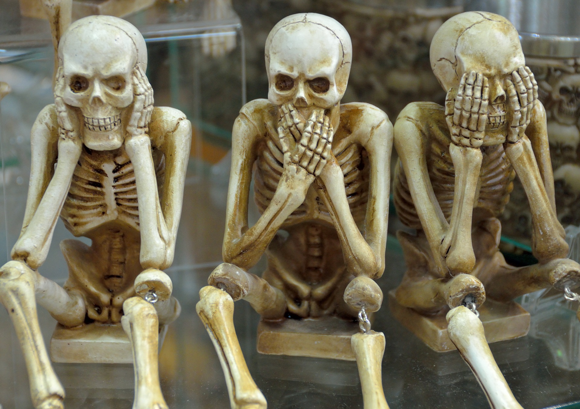
Our bones: strength, flexibility and…fractals!
The combined hardness and toughness of bone cannot be explained by the mere mixture of proteins and calcium phosphate mineral. To solve this conundrum, a deeper insight into the structure of this remarkable material is required. Using advanced three-dimensional nanoscale imaging of the mineral in human bone, we highlighted the importance of its structural organization.
Each of us has a skeleton – that is ordinary. However, the bone of which a skeleton is made is anything but ordinary. From an engineering perspective bone provides an incredibly versatile support structure that performs remarkably well in a circus contortionist, a sumo wrestler, a toddler who is climbing up a bookshelf, and in the toddler’s grandfather leaping forward to prevent a bone-breaking fall. However, after fracture, bones can heal without traces, and they are preserved as fossils for millions of years. Until adulthood, bones increase in size a thousand-fold. But after all, bones consist of abundant bioavailable elements such as calcium, phosphorus, and carbon. The secret of bones is that they are both strong and flexible at the same time. In fact, many biological objects are strong and flexible: for example, wood, antlers, spider webs. Over millions of years of evolution, nature developed a strategy to obtain maximal functional efficacy from the cheapest inventory – something called “hierarchical organization”.
How do we study hierarchical materials? One important point is to keep track of the context of observations. With modern characterization methods, it is possible to image at high magnification and thus to identify details all the way down to the level of individual atoms. However, our high-resolution information only makes sense if we know about its hierarchical context. Like when using interactive maps, zooming into a beautiful remote town you want to visit won’t help you to get there unless you also carefully look at the roads that lead to that town. In materials science, an important tool used for precise sampling is a focused ion beam coupled to an electron microscope. The ion beam acts as a fine knife and cuts samples a few hundred atoms thick from an interesting area of a larger specimen. This smaller sample from a known location and context can be further used for high-resolution imaging. The second crucial factor in the research of hierarchical materials is that all such materials are three-dimensional. Therefore, in order to understand and correctly interpret imaging data, it must obtained in 3D. An important technique for this is electron tomography, where a tiny sample as thin as one thousandth of the thickness of a human hair is imaged in a sequence of hundreds of projections, each one being slightly rotated with respect to the other. These projection images can be digitally assembled into a 3D image of the object − this digital replica can then be viewed from any angle at any level and analyzed in detail.
In fact, our ability to combine these modern methods of sampling and imaging into a smooth workflow became possible only fairly recently. For more than 300 years bone is known to be hierarchically structured (the first description of 5 levels of bone hierarchical organization was made by the English surgeon and scientist Clopton Havers in 1691). When we obtained a tiny specimen of bone from a known location and of a known orientation, and when we looked at the bone crystallites with the aim of revealing their 3D shape and size, we discovered that these crystallites were already hierarchical at the nanometer scale (one billionth of a meter)! Moreover, the thin crystals were slightly curved, like delicate petals splaying away from the pedicle, or like loosely braided hair. Beyond this, the hierarchy repeated itself: curved and needle-shaped crystals were merging sideways into a platelet (like fingers and a palm), and several platelets were stacked together (like 2, 3 or 4 hands pressed together). These intricate hierarchical aggregates merged and further split.
When zooming out whilst recalling the context of these curved crystallites in bone, an intriguing picture emerges: entire bones such as a rib, or the collar bone, have a clearly twisted shape, as the grooves and ridges in long bones follow a slight screw-like course. Tiny capillaries, which nourish the bone, pierce the bone shaft with a delicate, screw-like trajectory. Around the capillaries, bone material is organized into osteons − concentric layers, reminiscent of leek stems. These layers are made of narrow bundles twisting around the central capillaries with a varying angle. The bundles are composed of mineralized collagen fibrils gently twisting around the bundle axis, like threads of a rope. Collagen is a fibrous protein that exists in triple helices, and the mineral crystallites, as our study showed, are also twisted. Thus, the organization of bones is self-similar, with the repeating pattern being a helix. Therefore we call this organization of bone fractal-like.
At all levels, the organization of bone components follows a helical motif. This makes bone both resilient and strong, as is essentially required in real life so that indeed the toddler could fall from the shelf, and the grandfather would drop his newspaper and leap towards the baby, and both would not sustain skeletal injuries. Nature endlessly reuses successful design strategies, and skeletons are not an exception: down to the bone, we are fractals.
Original Article:
N. Reznikov, M. Bilton, L. Lari, M. M. Stevens, R. Kroger, Fractal-like hierarchical organization of bone begins at the nanoscale. Science 360, (2018).Edited by:
Massimo Caine , Founder and Director
We thought you might like
Smaller, faster, more complex? Watching a phase transition with X-ray eyes
Sep 18, 2023 in Maths, Physics & Chemistry | 3 min read by Allan JohnsonWolves don’t go doggy in the Alps: two decades of genetic evidence
Jul 24, 2019 in Evolution & Behaviour | 3 min read by Christophe Dufresnes , Luca FumagalliCharting the immune landscape in brain cancers
Nov 27, 2020 in Health & Physiology | 4 min read by Klara Soukup , Johanna A. JoyceTidings from Before the Flood: how Artificial Intelligence Rediscovers Ancient Babylonian Texts
Sep 1, 2021 in Evolution & Behaviour | 3.5 min read by Shai Gordin , Avital Romach , Ethan FetayaMore from Maths, Physics & Chemistry
Natural products might just be our best weapon against antibiotic resistance
Apr 3, 2024 in Maths, Physics & Chemistry | 3.5 min read by Olivier Kirchhoffer , Jahn Nitschke , Jean-Luc WolfenderHeading underground with cold atoms
Dec 1, 2023 in Maths, Physics & Chemistry | 3 min read by Jamie Vovrosh , Sam Hedges , Farzad HayatiHolographic sound fields shape 3D matter without a touch
Nov 15, 2023 in Maths, Physics & Chemistry | 4 min read by Kai MeldeHow to make a kilonova: Finding a path for cosmic alchemy
Oct 25, 2023 in Maths, Physics & Chemistry | 3.5 min read by Noel Richardson , Clarissa PavaoSurfing the Waves of Quantum Matter in Warm Classical Seas
Oct 23, 2023 in Maths, Physics & Chemistry | 4 min read by Imran Saeed , Tsvi Tulsty , Hyuk Kyu PakEditor's picks
Trending now
Popular topics